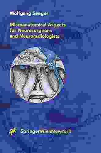
Microanatomical Aspects for Neurosurgeons and Neuroradiologists: A Comprehensive Guide

Microanatomy is the study of anatomical structures on a microscopic level. It is an essential field for neurosurgeons and neuroradiologists, as it provides a detailed understanding of the brain and spinal cord. This understanding is essential for the safe and effective diagnosis and treatment of neurological disorders.
5 out of 5
| Language | : | English |
| File size | : | 132998 KB |
| Text-to-Speech | : | Enabled |
| Screen Reader | : | Supported |
| Enhanced typesetting | : | Enabled |
| Print length | : | 531 pages |
| Paperback | : | 54 pages |
| Item Weight | : | 6.9 ounces |
| Dimensions | : | 8.5 x 0.14 x 11 inches |
The history of microanatomy can be traced back to the early days of microscopy. In the 16th century, Antonie van Leeuwenhoek used a microscope to observe living cells for the first time. In the 19th century, Camillo Golgi and Santiago Ramón y Cajal developed staining techniques that made it possible to visualize individual neurons and their connections. These techniques revolutionized our understanding of the nervous system and laid the foundation for the field of microanatomy.
Today, microanatomy is a highly specialized field that uses a variety of techniques to study the structure of the nervous system. These techniques include:
* Light microscopy: This technique uses visible light to visualize cells and tissues. It is a relatively simple and inexpensive technique, but it does not provide as much detail as other techniques. * Electron microscopy: This technique uses a beam of electrons to visualize cells and tissues. It provides much greater detail than light microscopy, but it is also more expensive and time-consuming. * Confocal microscopy: This technique uses a laser to scan a sample and create a three-dimensional image. It provides excellent detail and can be used to visualize live cells. * Magnetic resonance imaging (MRI): This technique uses a strong magnetic field and radio waves to create detailed images of the brain and spinal cord. It is a non-invasive technique that can be used to visualize both healthy and diseased tissue. * Computed tomography (CT): This technique uses X-rays to create detailed images of the brain and spinal cord. It is a non-invasive technique that can be used to visualize both healthy and diseased tissue.
Microanatomy has a wide range of applications in neurosurgery and neuroradiology. It is used to:
* Diagnose neurological disorders * Plan surgical procedures * Guide surgical interventions * Evaluate the results of surgery * Develop new treatments for neurological disorders
Microanatomy is a rapidly growing field that is constantly evolving. New techniques are being developed all the time, and our understanding of the nervous system is constantly expanding. This growth is driven by the need for better ways to diagnose and treat neurological disorders.
Microanatomy of the Brain
The brain is the most complex organ in the human body. It is responsible for controlling all of our thoughts, emotions, and movements. The brain is divided into two hemispheres, the left and right hemispheres. Each hemisphere is further divided into four lobes: the frontal lobe, parietal lobe, temporal lobe, and occipital lobe.
The frontal lobe is responsible for higher-level cognitive functions, such as planning, problem-solving, and decision-making. The parietal lobe is responsible for processing sensory information, such as touch, temperature, and pain. The temporal lobe is responsible for processing auditory information, such as speech and music. The occipital lobe is responsible for processing visual information.
The brain is also divided into a number of different regions, each of which has a specific function. These regions include the:
* Cerebral cortex: The cerebral cortex is the outer layer of the brain. It is responsible for higher-level cognitive functions, such as thinking, learning, and memory. * Basal ganglia: The basal ganglia are a group of structures located deep within the brain. They are responsible for controlling movement. * Thalamus: The thalamus is a structure located in the center of the brain. It serves as a relay station for sensory information. * Hypothalamus: The hypothalamus is a structure located at the base of the brain. It is responsible for regulating body temperature, hunger, and thirst. * Pituitary gland: The pituitary gland is a small gland located at the base of the brain. It produces hormones that regulate growth, development, and reproduction.
Microanatomy of the Spinal Cord
The spinal cord is a long, thin structure that runs from the brain down the back. It is responsible for transmitting messages between the brain and the rest of the body. The spinal cord is divided into 31 segments, each of which gives off a pair of spinal nerves.
The spinal cord is surrounded by a number of different structures, including the vertebral column, the meninges, and the cerebrospinal fluid. The vertebral column is a series of bones that protect the spinal cord. The meninges are three layers of connective tissue that surround the spinal cord and the cerebrospinal fluid is a clear liquid that fills the space between the meninges and the spinal cord.
The spinal cord is divided into four main regions:
* Cervical region: The cervical region is the upper region of the spinal cord. It gives off eight pairs of spinal nerves that innervate the neck, head, and upper limbs. * Thoracic region: The thoracic region is the middle region of the spinal cord. It gives off 12 pairs of spinal nerves that innervate the chest and abdomen. * Lumbar region: The lumbar region is the lower region of the spinal cord. It gives off five pairs of spinal nerves that innervate the lower back and lower limbs. * Sacral region: The sacral region is the lowest region of the spinal cord. It gives off five pairs of spinal nerves that innervate the pelvis and lower limbs.
Clinical Applications of Microanatomy
Microanatomy has a wide range of applications in neurosurgery and neuroradiology. These applications include:
* Diagnosis of neurological disorders: Microanatomy can be used to diagnose a variety of neurological disorders, including tumors, strokes, and dementias. * Planning surgical procedures: Microanatomy can be used to plan surgical procedures, such as brain surgery and spinal surgery. * Guiding surgical interventions: Microanatomy can be used to guide surgical interventions, such as biopsy and tumor resection. * Evaluating the results of surgery: Microanatomy can be used to evaluate the results of surgery, such as the extent of tumor removal and the presence of complications. * Developing new treatments for neurological disorders: Microanatomy can be used to develop new treatments for neurological disorders, such as gene therapy and stem cell therapy.
Microanatomy is a rapidly growing field that is constantly evolving. New techniques are being developed all the time, and our understanding of the nervous system is constantly expanding. This growth is driven by the need for better ways to diagnose and treat neurological disorders.
Microanatomy is an essential field for neurosurgeons and neuroradiologists. It provides a detailed understanding of the brain and spinal cord, which is essential for the safe and effective diagnosis and treatment of neurological disorders. Microanatomy is a rapidly growing field that is constantly evolving. New techniques are being developed all the time, and our understanding of the nervous system is constantly expanding. This growth is driven by the need for better ways to diagnose and treat neurological disorders.
5 out of 5
| Language | : | English |
| File size | : | 132998 KB |
| Text-to-Speech | : | Enabled |
| Screen Reader | : | Supported |
| Enhanced typesetting | : | Enabled |
| Print length | : | 531 pages |
| Paperback | : | 54 pages |
| Item Weight | : | 6.9 ounces |
| Dimensions | : | 8.5 x 0.14 x 11 inches |
Do you want to contribute by writing guest posts on this blog?
Please contact us and send us a resume of previous articles that you have written.
 Page
Page Text
Text Story
Story Genre
Genre Reader
 E-book
 Paragraph
 Sentence
 Bookmark
 Shelf
Shelf Bibliography
 Foreword
 Preface
 Synopsis
 Manuscript
 Codex
Codex Tome
Tome Bestseller
 Classics
 Narrative
 Biography
 Autobiography
 Reference
 Dictionary
 Thesaurus
 Resolution
 Catalog
 Card Catalog
 Stacks
 Archives
 Periodicals
 Study
Study Research
 Rare Books
 Special Collections
 Literacy
 Study Group
 Reading List
 Theory
 Textbooks
 Iain Lawrence
 James Brown
 Polly Pallister Wilkins
 Rajat Paharia
 Tim Browning
 Rick Wormeli
 Zess
Zess Nicki Bell
 Patrick Mcnamara
 Nathaniel Hawthorne
 Sheila Christensen
 Lynne A Weikart
 Cynthia Woolf
 A L Noble
 Sophie Claire
 David Oliver
 Leonard Pitts
 Karen Cushman
 Judith Frege
 Christie Barlow
Light bulbAdvertise smarter! Our strategic ad space ensures maximum exposure. Reserve your spot today!

 Arthur C. ClarkeThe Enchanting Language of Flowers: A Guide to the English Alphabet and Basic...
 José MartíFollow ·19.5k
 Ralph TurnerFollow ·4.4k
 Joseph ConradFollow ·10.1k
 Bill GrantFollow ·9.8k
 Jim CoxFollow ·2.5k
 Arthur C. ClarkeFollow ·12k
 Ron BlairFollow ·3k
 Leon FosterFollow ·17.4k

 Al Foster
How To Breathe Underwater: Unlocking the Secrets of...
: Embracing the...

 Ian Mitchell
The Laws of Gravity: A Literary Journey into the...
Lisa Ann Gallagher's...

 Francis Turner
Christmas Solos For Beginning Viola: A Detailed Guide for...
Christmas is a time for...

 Jamal Blair
Define Humanistic Psychology Forms Of Communication...
Humanistic...

 Morris Carter
Judgment in Berlin: Unraveling the Intrigue of an...
"Judgment in Berlin" is a gripping...
5 out of 5
| Language | : | English |
| File size | : | 132998 KB |
| Text-to-Speech | : | Enabled |
| Screen Reader | : | Supported |
| Enhanced typesetting | : | Enabled |
| Print length | : | 531 pages |
| Paperback | : | 54 pages |
| Item Weight | : | 6.9 ounces |
| Dimensions | : | 8.5 x 0.14 x 11 inches |


